
Neuroimaging is a fascinating field of medical modern science. It involves using various imaging techniques to visualize the structure and function of the brain and spinal cord.
Structural Imaging
This technique provides images of the brain’s anatomy.
Examples are:
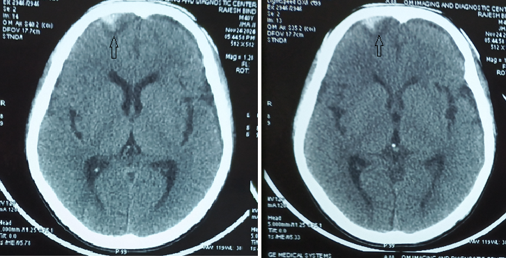
Computed Tomography (CT): Uses X-rays to create detailed images of the brain. Its variants include CT angiography (CTA); perfusion CT (pCT) and dual-energy CT.
Magnetic Resonance Imaging (MRI): Uses very strong magnetic fields to produce high-resolution images of the brain’s structure. Its variants include MR angiography (MRA); MR vessel wall imaging; MR neurography (MRN).
Functional Imaging
Functional techniques measure brain activity and function. Examples are:
Functional MRI (fMRI); MR spectroscopy (MRS); diffusion-weighted MR imaging (DWI); diffusion tensor MR imaging (DTI); susceptibility-weighted MR imaging (SWI); arterial spin label imaging (ASL); and perfusion MRI (pMRI); Positron Emission Tomography (PET).
Interventional techniques
There are also a number of interventional neuroradiologic techniques such as catheter embolization, stent retrieval thrombectomy, aneurysm coiling and stenting.
Imaging techniques for spine
Numerous techniques are also employed to diagnose spine disorders such as CT myelography, fluoroscopy and CT-guided spine interventional procedures for pain and oncology, including radiofrequency and cold ablation and image guided blood patches.
Angiography techniques
Multidetector computed tomographic angiography (MDCTA)and gadolinium enhanced magnetic resonance angiography(MRA) techniques are replacing catheter-based angiography, which is now reserved for patients in whom small-vessel detail is essential for diagnosis or for whom concurrent interventional therapy is planned.
MRI
MRI is more sensitive than CT for the detection of lesions affecting the peripheral and central nervous system (CNS). Since CT can be done more quickly, it is a good choice for uncooperative patients, or those with suspected acute stroke, hemorrhage, and acute intracranial or spinal trauma.
CT is more sensitive than MRI for visualizing fine bony details and so it is an appropriate initial imaging technique for assessing conductive hearing loss, and lesions affecting the bony parts of skull and spine. But MRI is more informative regarding bone marrow infiltrative processes that can be difficult to detect on CT.
Diffusion-weighted MRI
Diffusion MRI is the most sensitive technique for detecting acute ischemic stroke of the brain or spinal cord. It is also useful in the detection of encephalitis, abscess, Creutzfeldt-Jacob disease, cerebral tumors, and acute demyelinating lesions.
Leave a Reply